
graticule scale is 20 mm per small division
Selected-area diffraction of a disordered polycrystalline aggregate
A. van Donkelaar, CSIRO Health Sciences
SYNOPSIS: Use of a narrow beam permits the selected-area diffraction technique to be used on a protein-crystal aggregate to isolate a good single crystal for structure refinement analysis.
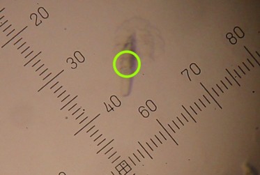
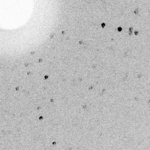
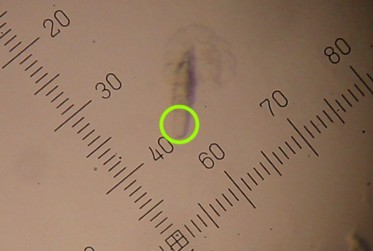
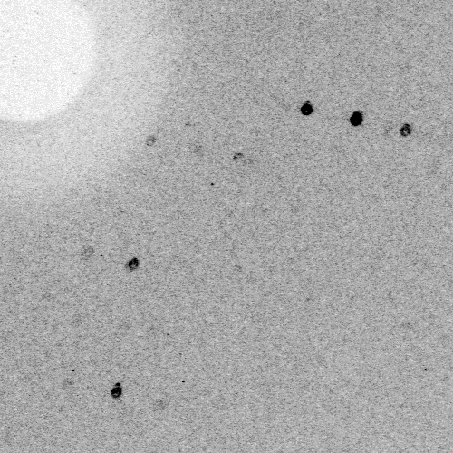
The narrow focussed beam produced by the PX50 optical system permits the selected-area diffraction technique to be applied to polycrystalline aggregates in which small single protein crystals can be isolated within the 0.1mm beam diameter over a feasible oscillation range. An example of this situation, in which crystallisation of a neuraminidase mutant yielded a single polycrystalline specimen, initially yielded no useful diffraction data when irradiated centrally (see figures). The observation of a single-crystal protrusion from the aggregate prompted the attempt to orient the aggregate to isolate the single-crystal in the beam, leading to a successful structure refinement to 3Å from a crystal possessing a relatively large unit-cell volume despite an irradiated crystal volume of less than 100x40x30 mm.
Selected-area diffraction patterns from adjacent areas of a polycrystalline aggregate of neuraminidase-mutant crystals. Circles show beam position (after specimen was mounted and frozen in cryostream) corresponding to observed diffraction patterns of 40 mins exposure at 300mm camera length.
Crystal data: P2 monoclinic structure, a=315.4Å, b=67.6Å, c=92.6Å, a=90°, b=92.6°, g=90°.
 |
 |
|
Circle indicates beam size and position graticule scale is 20 mm per small division |
 |
 |
|
Circle indicates beam size and position graticule scale is 20 mm per small division |
(c) Copyright AXCO 2004